Building the virtual molecular layer for tissue biology
Aurora predicts spatial gene expression from routine H&E images, helping researchers explore molecular patterns across samples, cohorts and disease.
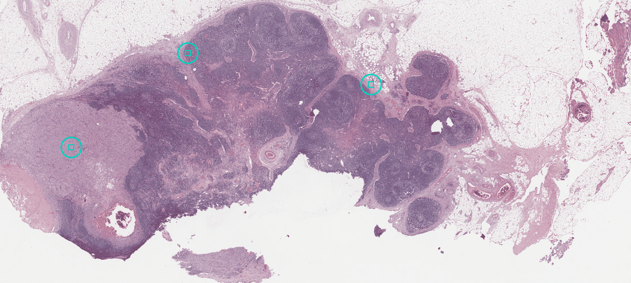
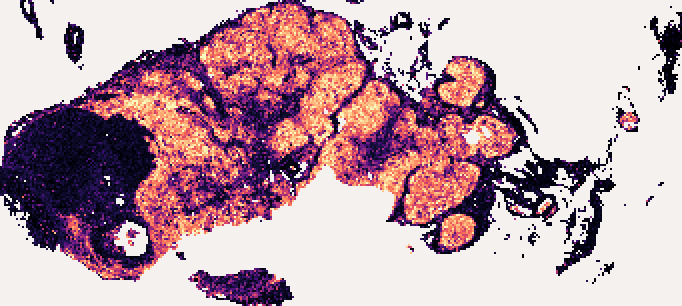
MS4A1 over the 24,308 retained tissue tiles in that region. Inspect the slide and both gene maps.What Aurora helps you do
- Past tissue. Future questions. A cohort assembled years ago can meet a question that emerged yesterday.
- Find patterns at cohort scale. Explore virtual molecular variation across samples and biological groups.
- Prioritise what to pursue. Focus validation and experimental effort on the signals, samples and hypotheses that matter most.
Explore research applications on the website, or explore one real slide here.
From existing tissue to a virtual molecular view
Virtual spatial transcriptomics is predicted, spatially resolved gene expression from H&E images. Aurora returns it on the slide's own coordinates as one transcriptome-wide matrix. No spatial assay is run.
Choose the workflow that fits your research
The tissue is already here. A new layer of biology is within reach.
-
Upload your slides
The simple path
Aurora Direct takes an H&E image and an email address, with no code and no account. Submit images for analysis. From code, install and prepare a slide, or compare the two routes.
-
Keep your slides with you
When images must stay in your environment
Run DeepSpot-H on your own machine and send the numbers it produces. Run DeepSpot-H locally.
Analysis is for academic and non-profit research. Commercial evaluation and use are governed by a written agreement with Aurora. Discuss commercial access.
The models behind a prediction
Two models make each prediction. DeepSpot-H reads the tissue's structure in each part of the slide, so DeepSpot-M can predict spatial gene expression there. Learn about DeepSpot-H or learn about DeepSpot-M.